A pair of atria, an aorta, a tricuspid valve, a pulmonary vein, the superior vena cava. All this—the human heart—is just a minuscule cylinder at first.
And that’s pretty wild.
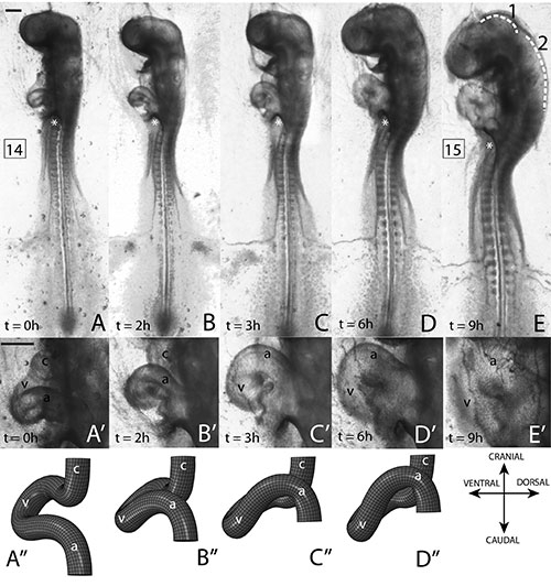
S-looping heart development in chicken embryo (By Sarah Bradner '14)
“Initially, the embryonic heart forms as a straight tube, like a garden hose. To become the complex mature organ, it twists and bends as a baby grows,” Ashok Ramasubramanian explained, curling a string from his winter hat in demonstration. “But there are no fingers inside; the heart has to bend by itself—a process called looping.”
“Certain genes change its shape, but there are also forces,” he continued. “Consider that you go to California and your head is a gene. If your head falls off you won’t go, but you can’t just say your head is all that gets you there. Other forces, like a plane, are involved. It’s these—the mechanisms of gene action—we study to see how the heart actually twists.”
Ramasubramanian and his colleagues analyze chicken embryos (their development is similar to humans’) using computer models and an atomic force microscope. Both help identify forces acting on the chick heart, which is only 1 mm long with a tube circumference of 400 microns in the s-looping stage they’re studying.
During this critical period, occurring in the first 48 to 56 hours of a chick’s 21-day incubation, groundwork is laid for the basic cardiac shape—two atria at the top and two ventricles at the bottom.
“Many babies are born with heart abnormalities, but most of these conditions we can’t treat in utero, partly because cardiac development is poorly understood,” Ramasubramanian said. “We study this with the hope of understanding.”
The team’s work is funded by an NIH Academic Research Enhancement Award.